Publications
Filter by type:
Tackling standardization in fluorescence molecular imaging
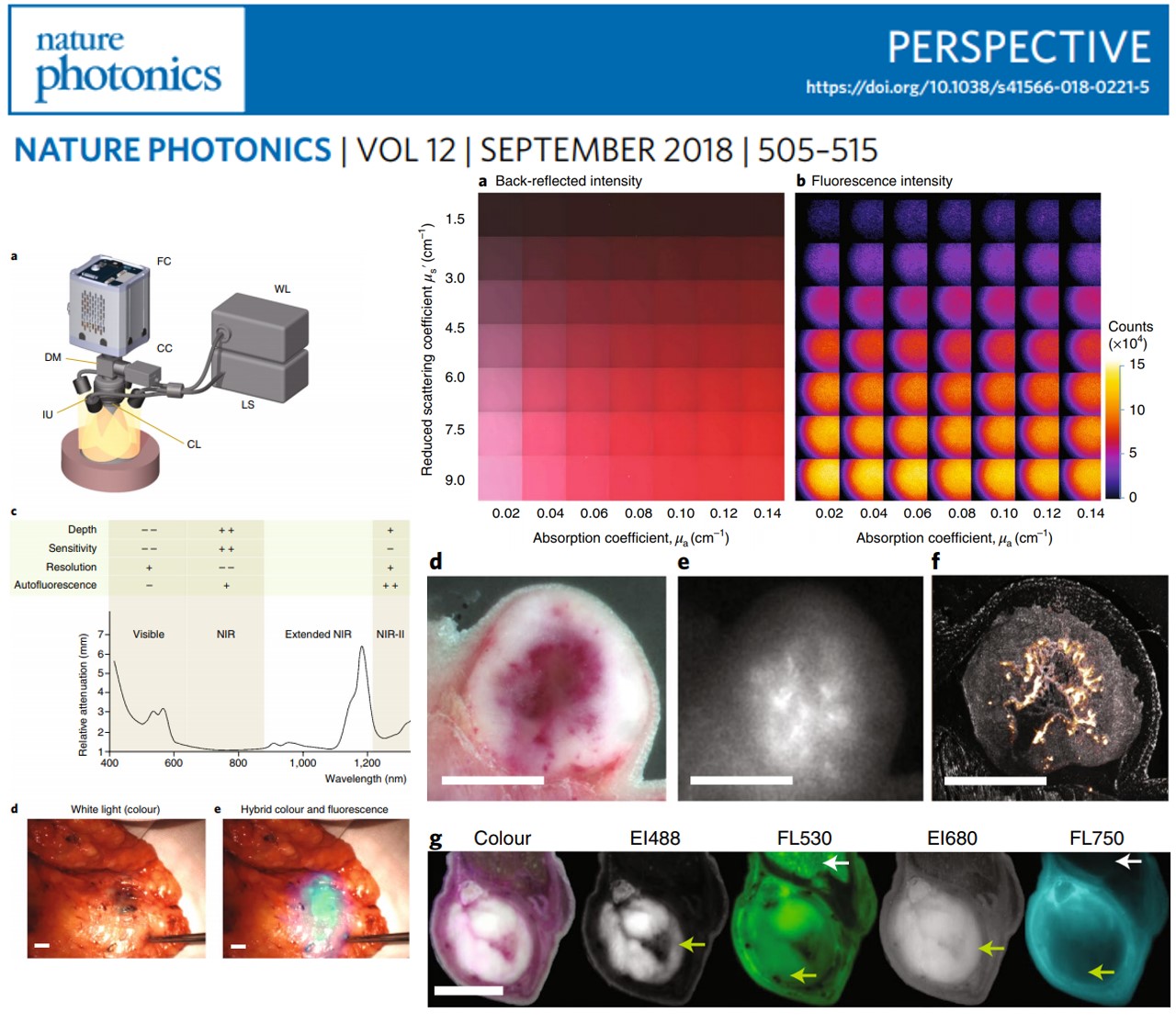
The emerging clinical use of targeted fluorescent agents heralds a shift in intraoperative imaging practices that overcome the limitations of human vision. However, in contrast to established radiological methods, no appropriate performance specifications and standards have been established in fluorescence molecular imaging. Moreover, the dependence of fluorescence signals on many experimental parameters and the use of wavelengths ranging from the visible to short-wave infrared (400–1,700 nm) complicate quality control in fluorescence molecular imaging. Here, we discuss the experimental parameters that critically affect fluorescence molecular imaging accuracy, and introduce the concept of high-fidelity fluorescence imaging as a means for ensuring reliable reproduction of fluorescence biodistribution in tissue.
Nature Photonics, Volume 12, pages505–515 (2018),2018
Threshold Analysis and Biodistribution of Fluorescently Labeled Bevacizumab in Human Breast Cancer
In vivo tumor labeling with fluorescent agents may assist endoscopic and surgical guidance for cancer therapy as well as create opportunities to directly observe cancer biology in patients. However, malignant and non-malignant tissues are usually distinguished on fluorescence images by applying empirically determined fluorescence intensity thresholds. Here we report the development of fSTREAM, a set of analytic methods designed to streamline the analysis of surgically excised breast tissues by collecting and statistically processing hybrid multi-scale fluorescence, color, and histology readouts toward precision fluorescence imaging. fSTREAM addresses core questions of how to relate fluorescence intensity to tumor tissue and how to quantitatively assign a normalized threshold that sufficiently differentiates tumor tissue from healthy tissue. Using fSTREAM we assessed human breast tumors stained in vivo with fluorescent bevacizumab at microdose levels Showing that detection of such levels is achievable, we validated fSTREAM for high-resolution mapping of the spatial pattern of labeled antibody and its relation to the underlying cancer pathophysiology and tumor border on a per patient basis. We demonstrated a 98% sensitivity and 79% specificity when using labelled bevacizumab to outline the tumor mass. Overall, our results illustrate a quantitative approach to relate fluorescence signals to malignant tissues and improve the theranostic application of fluorescence molecular imaging.
Cancer Research, 2017 Feb 1;77(3):623-631,2017
NeuBtracker—imaging neurobehavioral dynamics in freely behaving fish
A long-standing objective in neuroscience has been to image distributed neuronal activity in freely behaving animals. Here we introduce NeuBtracker, a tracking microscope for simultaneous imaging of neuronal activity and behavior of freely swimming fluorescent reporter fish. We showcase the value of NeuBtracker for screening neurostimulants with respect to their combined neuronal and behavioral effects and for determining spontaneous and stimulus-induced spatiotemporal patterns of neuronal activation during naturalistic behavior.
Nat. Methods, vol. 14, no. 11, pp. 1079–1082, 2017,2017
Calcium Sensor for Photoacoustic Imaging
We introduce a selective and cell-permeable calcium sensor for photoacoustics (CaSPA), a versatile imaging technique that allows for fast volumetric mapping of photoabsorbing molecules with deep tissue penetration. To optimize for Ca2+-dependent photoacoustic signal changes, we synthesized a selective metallochromic sensor with high extinction coefficient, low quantum yield, and high photobleaching resistance. Micromolar concentrations of Ca2+ lead to a robust blueshift of the absorbance of CaSPA, which translated into an accompanying decrease of the peak photoacoustic signal. The acetoxymethyl esterified sensor variant was readily taken up by cells without toxic effects and thus allowed us for the first time to perform live imaging of Ca2+ fluxes in genetically unmodified cells and heart organoids as well as in zebrafish larval brain via combined fluorescence and photoacoustic imaging.
In JACS,
2017
High-Resolution Multispectral Optoacoustic Tomography of the Vascularization and Constitutive Hypoxemia of Cancerous Tumors
Diversity of the design and alignment of illumination and ultrasonic transducers empower the fine scalability and versatility of optoacoustic imaging. In this study, we implement an innovative high-resolution optoacoustic mesoscopy for imaging the vasculature and tissue oxygenation within subcutaneous and orthotopic cancerous implants of mice in vivo through acquisition of tomographic projections over 180° at a central frequency of 24 MHz. High-resolution volumetric imaging was combined with multispectral functional measurements to resolve the exquisite inner structure and vascularization of the entire tumor mass using endogenous and exogenous optoacoustic contrast. Evidence is presented for constitutive hypoxemia within the carcinogenic tissue through analysis of the hemoglobin absorption spectra and distribution. Morphometric readouts obtained with optoacoustic mesoscopy have been verified with high-resolution ultramicroscopic studies. The findings described herein greatly extend the applications of optoacoustic mesoscopy toward structural and multispectral functional measurements of the vascularization and hemodynamics within solid tumors in vivo and are of major relevance to basic and preclinical oncological studies in small animal models.
In Neoplasia,
2016
Eigenspectra optoacoustic tomography achieves quantitative blood oxygenation imaging deep in tissues
Light propagating in tissue attains a spectrum that varies with location due to wavelength-dependent fluence attenuation, an effect that causes spectral corruption. Spectral corruption has limited the quantification accuracy of optical and optoacoustic spectroscopic methods, and impeded the goal of imaging blood oxygen saturation (sO2) deep in tissues; a critical goal for the assessment of oxygenation in physiological processes and disease. Here we describe light fluence in the spectral domain and introduce eigenspectra multispectral optoacoustic tomography (eMSOT) to account for wavelength-dependent light attenuation, and estimate blood sO2 within deep tissue. We validate eMSOT in simulations, phantoms and animal measurements and spatially resolve sO2 in muscle and tumours, validating our measurements with histology data. eMSOT shows substantial sO2 accuracy enhancement over previous optoacoustic methods, potentially serving as a valuable tool for imaging tissue pathophysiology.
In NatCom,
2016
Uniqueness in multispectral constant-wave epi-illumination imaging
Multispectral tissue imaging based on optical cameras and continuous-wave tissue illumination is commonly used in medicine and biology. Surprisingly, there is a characteristic absence of a critical look at the quantities that can be uniquely characterized from optically diffuse matter by multispectral imaging. Here, we investigate the fundamental question of uniqueness in epi-illumination measurements from turbid media obtained at multiple wavelengths. By utilizing an analytical model, tissue-mimicking phantoms, and an in vivo imaging experiment we show that independent of the bands employed, spectral measurements cannot uniquely retrieve absorption and scattering coefficients. We also establish that it is, nevertheless, possible to uniquely quantify oxygen saturation and the Mie scattering power—a previously undocumented uniqueness condition.
In Opt.Lett.,
2016
Pushing the Optical Imaging Limits of Cancer with Multi-Frequency-Band Raster-Scan Optoacoustic Mesoscopy (RSOM)
Angiogenesis is a central cancer hallmark, necessary for supporting tumor growth and metastasis. In vivo imaging of angiogenesis is commonly applied, to understand dynamic processes in cancer development and treatment strategies. However, most radiological modalities today assess angiogenesis based on indirect mechanisms, such as the rate of contrast enhancement after contrast agent administration. We studied the performance of raster-scan optoacoustic mesoscopy (RSOM), to directly reveal the vascular network supporting melanoma growth in vivo, at 50 MHz and 100 MHz, through several millimeters of tumor depth. After comparing the performance at each frequency, we recorded, for the first time, high-resolution images of melanin tumor vasculature development in vivo, over a period of several days. Image validation was provided by means of cryo-slice sections of the same tumor after sacrificing the mice. We show how optoacoustic (photoacoustic) mesoscopy reveals a potentially powerful look into tumor angiogenesis, with properties and features that are markedly different than other radiological modalities. This will facilitate a better understanding of tumor’s angiogenesis, and the evaluation of treatment strategies.
In Neoplasia,
2016
Calcium neuroimaging in behaving zebrafish larvae using a turn-key light field camera
Reconstructing a three-dimensional scene from multiple simultaneously acquired perspectives (the light field) is an elegant scanless imaging concept that can exceed the temporal resolution of currently available scanning-based imaging methods for capturing fast cellular processes. We tested the performance of commercially available light field cameras on a fluorescent microscopy setup for monitoring calcium activity in the brain of awake and behaving reporter zebrafish larvae. The plenoptic imaging system could volumetrically resolve diverse neuronal response profiles throughout the zebrafish brain upon stimulation with an aversive odorant. Behavioral responses of the reporter fish could be captured simultaneously together with depth-resolved neuronal activity. Overall, our assessment showed that with some optimizations for fluorescence microscopy applications, commercial light field cameras have the potential of becoming an attractive alternative to custom-built systems to accelerate molecular imaging research on cellular dynamics.
J. of Biomedical Optics, 20(9), 096009 (2015).,2015
A publication title, such as title of a paper
Optical mesoscopy extends the capabilities of biological visualization beyond the limited penetration depth achieved by microscopy. However, imaging of opaque organisms or tissues larger than a few hundred micrometers requires invasive tissue sectioning or chemical treatment of the specimen for clearing photon scattering, an invasive process that is regardless limited with depth. We developed previously unreported broadband optoacoustic mesoscopy as a tomographic modality to enable imaging of optical contrast through several millimeters of tissue, without the need for chemical treatment of tissues. We show that the unique combination of three-dimensional projections over a broad 500 kHz-40 MHz frequency range combined with multi-wavelength illumination is necessary to render broadband multispectral optoacoustic mesoscopy (2B-MSOM) superior to previous optical or optoacoustic mesoscopy implementations.
In Biomed. Opt. Express,
2015
Quantitative detection of drug dose and spatial distribution in the lung revealed by Cryoslicing Imaging.
Administration of drugs via inhalation is an attractive route for pulmonary and systemic drug delivery. The therapeutic outcome of inhalation therapy depends not only on the dose of the lung-delivered drug, but also on its bioactivity and regional distribution. Fluorescence imaging has the potential to monitor these aspects already during preclinical development of inhaled drugs, but quantitative methods of analysis are lacking. In this proof-of-concept study, we demonstrate that Cryoslicing Imaging allows for 3D quantitative fluorescence imaging on ex vivo murine lungs. Known amounts of fluorescent substance (nanoparticles or fluorophore–drug conjugate) were instilled in the lungs of mice. The excised lungs were measured by Cryoslicing Imaging. Herein, white light and fluorescence images are obtained from the face of a gradually sliced frozen organ block. A quantitative representation of the fluorescence intensity throughout the lung was inferred from the images by accounting for instrument noise, tissue autofluorescence and out-of-plane fluorescence. Importantly, the out-of-plane fluorescence correction is based on the experimentally determined effective light attenuation coefficient of frozen murine lung tissue (10.0 ± 0.6 cm−1 at 716 nm). The linear correlation between pulmonary total fluorescence intensity and pulmonary fluorophore dose indicates the validity of this method and allows direct fluorophore dose assessment. The pulmonary dose of a fluorescence-labeled drug (FcγR-Alexa750) could be assessed with an estimated accuracy of 9% and the limit of detection in ng regime. Hence, Cryoslicing Imaging can be used for quantitative assessment of dose and 3D distribution of fluorescence-labeled drugs or drug carriers in the lungs of mice.
In J.Pharm.Biomed.Anal.,
2015
Serial sectioning and multispectral imaging system for versatile biomedical applications
Serial sectioning combined with microscopy provides high resolution volumetric data to complement in-vivo imaging modalities and aid ex-vivo diagnostics. We describe the design of a fully-automated cryomicrotome combined with a multispectral reflection and fluorescence imaging system that enables high-throughput analyses of biological specimens with a large field of view and cellular resolution while keeping the manufacturing and running costs low. We show the performance of the system for representative applications in high-resolution volumetric imaging of reporter animals and multispectral tissue analysis. Furthermore, we demonstrate the versatility of the economical imaging system in applications such as in vivo epifluorescence imaging, histology slide scanning, cell counting and gel electrophoresis documentation.
In IEEE-ISBI Proc.,
2014
Steady-state total diffuse reflectance with an exponential decaying source
The increasing preclinical and clinical utilization of digital cameras for photographic measurements of tissue conditions motivates the study of reflectance measurements obtained with planar illumination. We examine herein a formula that models the total diffuse reflectance measured from a semi-infinite medium using an exponentially decaying source, assuming continuous plane wave epi-illumination. The model is validated with experimental reflectance measurements from tissue mimicking phantoms. The need for adjusting the blood absorption spectrum due to pigment packaging is discussed along with the potential applications of the proposed formulation.
In Opt.Lett.,
2014
Robust overlay schemes for the fusion of fluorescence and color channels in biological imaging
Molecular fluorescence imaging is a commonly used method in various biomedical fields and is undergoing rapid translation toward clinical applications. Color images are commonly superimposed with fluorescence measurements to provide orientation, anatomical information, and molecular tissue properties in a single image. New adaptive methods that produce a more robust composite image than conventional lime green alpha blending are presented and demonstrated herein. Moreover, visualization through temporal changes is showcased as an alternative for real-time imaging systems.
In JBO,
2014
Towards clinically translatable NIR fluorescence molecular guidance for colonoscopy
White-light surveillance colonoscopy is the standard of care for the detection and removal of premalignant lesions to prevent colorectal cancer, and the main screening recommendation following treatment for recurrence detection. However, it lacks sufficient diagnostic yield, exhibits unacceptable adenoma miss-rates and is not capable of revealing functional and morphological information of the detected lesions. Fluorescence molecular guidance in the near-infrared (NIR) is expected to have outstanding relevance regarding early lesion detection and heterogeneity characterization within and among lesions in these interventional procedures. Thereby, superficial and sub-surface tissue biomarkers can be optimally visualized due to a minimization of tissue attenuation and autofluorescence by comparison with the visible, which simultaneously enhance tissue penetration and assure minimal background. At present, this potential is challenged by the difficulty associated with the clinical propagation of disease-specific contrast agents and the absence of a commercially available endoscope that is capable of acquiring wide-field, NIR fluorescence at video-rates. We propose two alternative flexible endoscopic fluorescence imaging methods, each based on a CE certified commercial, clinical grade endoscope, and the employment of an approved monoclonal antibody labeled with a clinically applicable NIR fluorophore. Pre-clinical validation of these two strategies that aim at bridging NIR fluorescence molecular guidance to clinical translation is demonstrated in this study.
In Biomed Opt Express,
2014
Three-dimensional imaging of whole mouse models: comparing nondestructive X-ray phase-contrast micro-CT with cryotome-based planar epi-illumination imaging
In this study, we compare two evolving techniques for obtaining high-resolution 3D anatomical data of a mouse specimen. On the one hand, we investigate cryotome-based planar epi-illumination imaging (cryo-imaging). On the other hand, we examine X-ray phase-contrast micro-computed tomography (micro-CT) using synchrotron radiation. Cryo-imaging is a technique in which an electron multiplying charge coupled camera takes images of a cryo-frozen specimen during the sectioning process. Subsequent image alignment and virtual stacking result in volumetric data. X-ray phase-contrast imaging is based on the minute refraction of X-rays inside the specimen and features higher soft-tissue contrast than conventional, attenuation-based micro-CT. To explore the potential of both techniques for studying whole mouse disease models, one mouse specimen was imaged using both techniques. Obtained data are compared visually and quantitatively, specifically with regard to the visibility of fine anatomical details. Internal structure of the mouse specimen is visible in great detail with both techniques and the study shows in particular that soft-tissue contrast is strongly enhanced in the X-ray phase images compared to the attenuation-based images. This identifies phase-contrast micro-CT as a powerful tool for the study of small animal disease models.
In J.Microsc.,
2014
Vaccinia virus-mediated melanin production allows MR and optoacoustic deep tissue imaging and laser-induced thermotherapy of cancer
We reported earlier the delivery of antiangiogenic single chain antibodies by using oncolytic vaccinia virus strains to enhance their therapeutic efficacy. Here, we provide evidence that gene-evoked production of melanin can be used as a therapeutic and diagnostic mediator, as exemplified by insertion of only one or two genes into the genome of an oncolytic vaccinia virus strain. We found that produced melanin is an excellent reporter for optical imaging without addition of substrate. Melanin production also facilitated deep tissue optoacoustic imaging as well as MRI. In addition, melanin was shown to be a suitable target for laser-induced thermotherapy and enhanced oncolytic viral therapy. In conclusion, melanin as a mediator for thermotherapy and reporter for different imaging modalities may soon become a versatile alternative to replace fluorescent proteins also in other biological systems. After ongoing extensive preclinical studies, melanin overproducing oncolytic virus strains might be used in clinical trials in patients with cancer.
In PNAS,
2013


